
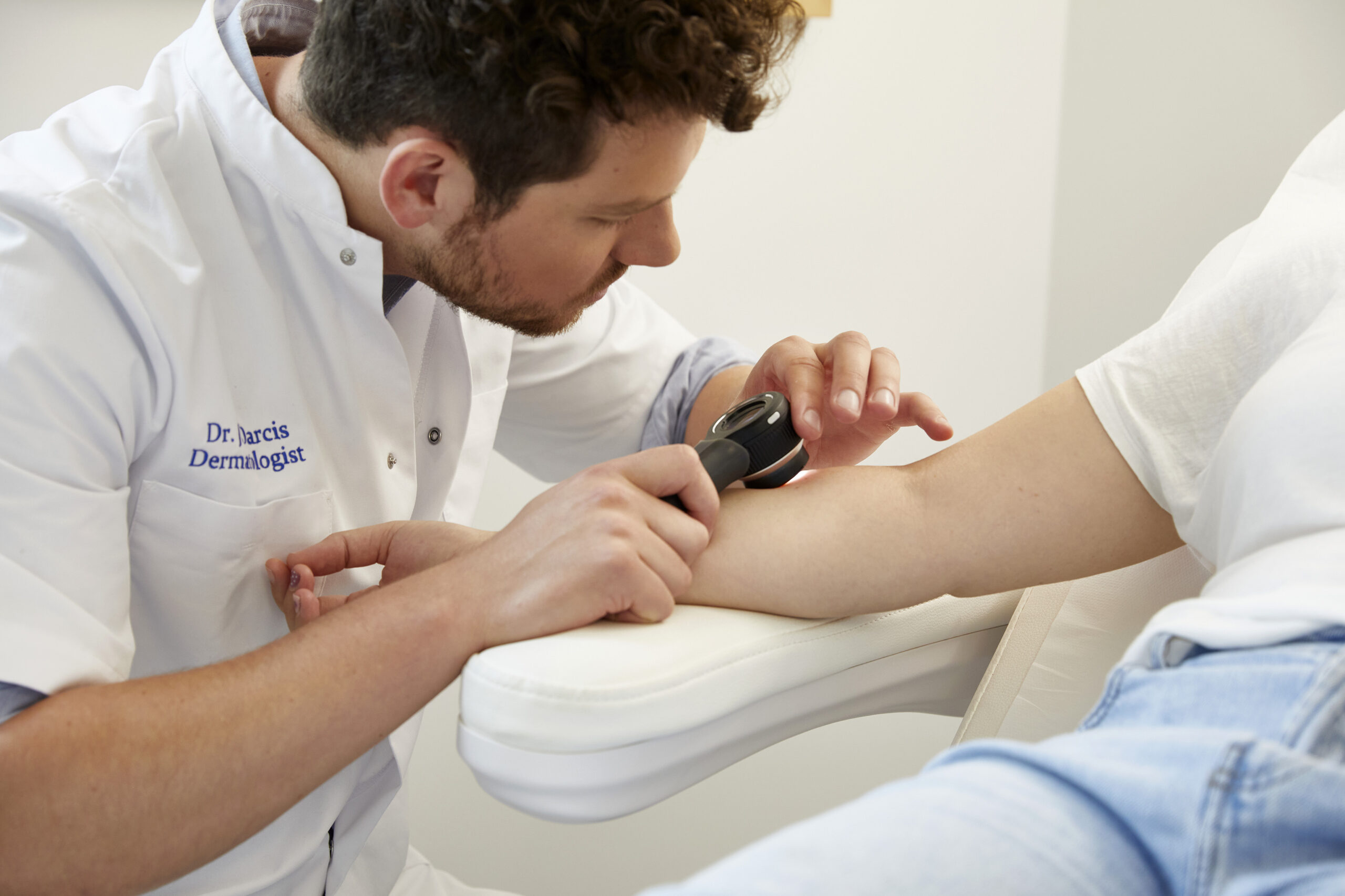
Dermoscopy
Dermoscopy or epiluminescent microscopy is the examination of a skin lesion with a dermatoscope, a magnifying glass using polarised and non-polarised light.
It provides additional information to the clinical examination and it allows to make diagnosis and avoid an unnecessary procedure. Even if in some cases it is always safer to proceed with surgical removal of the skin lesion, followed by complete microscopic examination.
We use the dermoscopy to analyse moles but also to diagnose other benign or malignant skin lesions (verruca seborrheica, angiokeratoma, basal cell carcinoma, melanoma,..)
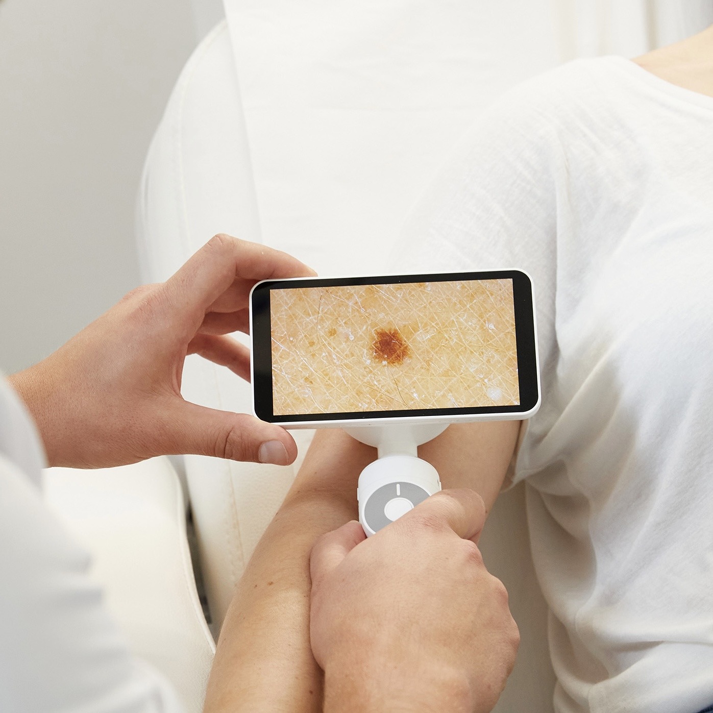
Digital mole mapping
We use the Barco Demetra Digital Dermoscopy system.
This is a digitalised mapping system of images of moles on the body using overview pictures of body parts, close up pictures and dermoscopic images.
Moles that need monitoring are digitalised during the clinical examinations, and re-evaluated in follow up visits.
It is an fast and effective way to monitor moles not only clinically but also with photographic and dermoscopic imaging, allowing to see minor changes that would not be visible with the naked eye.
Full body mole mapping is available on request (additional charges may apply).